How to Disrupt Vitamin B2’s Protective Shield to Induce Ferroptosis in Cancer Cells
Introduction
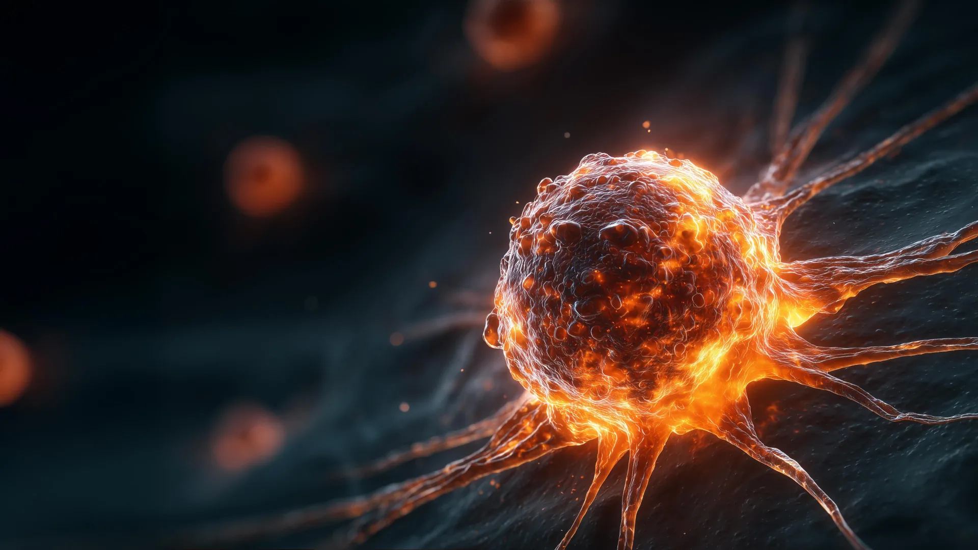
Recent scientific discoveries have unveiled an unexpected role for vitamin B2 (riboflavin) in cancer biology: it can help cancer cells survive by protecting them from a specialized form of programmed cell death known as ferroptosis. Ferroptosis is a promising anticancer mechanism, but some tumors evade it by using vitamin B2 to reinforce a cellular shield. In laboratory experiments, researchers found that a natural compound called roseoflavin—a close mimic of vitamin B2—can break down that shield and trigger cancer cell death. This guide walks you through the steps to replicate these findings, understand the underlying mechanisms, and explore potential therapeutic applications.
What You Need
- Cancer cell lines (e.g., those known to be sensitive to ferroptosis, such as certain lung, breast, or melanoma cells)
- Cell culture medium (DMEM or RPMI-1640 supplemented with 10% fetal bovine serum and antibiotics)
- Vitamin B2 (riboflavin) – stock solution (e.g., 10 mM in DMSO or water)
- Roseoflavin – a vitamin B2 analog (commercially available or synthesized; stock solution in DMSO)
- Ferroptosis inducers and inhibitors (optional for controls, e.g., erastin, RSL3, ferrostatin-1)
- Cell viability assay kit (e.g., MTT, CCK-8, or CellTiter-Glo)
- Lipid peroxidation detection reagents (e.g., C11-BODIPY 581/591 or TBARS assay kit)
- Flow cytometer or fluorescence microscope
- Standard lab equipment: CO₂ incubator, biosafety cabinet, centrifuge, pipettes, multiwell plates
Step-by-Step Instructions
Step 1: Understand the Role of Ferroptosis and Vitamin B2
Before starting experiments, it’s crucial to grasp the core concept: ferroptosis is an iron-dependent form of cell death driven by lipid peroxidation. Cells have natural defense mechanisms, including a system that uses vitamin B2 (as flavin cofactors) to reduce lipid peroxides and prevent cell death. In cancer cells, this vitamin B2–dependent shield can be overactive, making them resistant to ferroptosis-based therapies. Roseoflavin competes with vitamin B2 for binding to flavin enzymes, thereby disrupting the shield and sensitizing cells to ferroptosis.
Step 2: Culture Cancer Cells
Grow your chosen cancer cell line in appropriate culture medium at 37°C with 5% CO₂. Maintain cells in exponential growth phase. For experiments, seed cells in 96-well plates (for viability assays) or 6-well plates (for flow cytometry) at a density that allows ~80% confluency at the time of treatment—typically 5,000–10,000 cells per well for 96-well plates.
Step 3: Confirm Baseline Sensitivity to Ferroptosis
To establish a reliable model, first treat cells with a known ferroptosis inducer (e.g., 1–10 µM erastin or 0.1–1 µM RSL3) for 24–48 hours. Measure cell viability using an MTT or equivalent assay. Confirm that without vitamin B2 interference, cells die via ferroptosis (rescued by ferrostatin-1 or lipophilic antioxidants). This step validates your cell line and assay conditions.
Step 4: Expose Cells to Vitamin B2 to Enhance the Protective Shield
Pretreat cells with increasing concentrations of vitamin B2 (e.g., 10–100 µM) for 24 hours. Then, add a ferroptosis inducer (same concentration as Step 3). After 24–48 hours, measure cell viability. You should observe that vitamin B2 pretreatment significantly reduces ferroptosis-induced cell death—confirming its protective role.
Step 5: Apply Roseoflavin to Disrupt the Shield
Now introduce roseoflavin as a competitor. In parallel sets of wells, add roseoflavin at concentrations ranging from 10–100 µM (or equimolar to vitamin B2) along with vitamin B2 and the ferroptosis inducer. Incubate for 24–48 hours. Roseoflavin should outcompete vitamin B2 for binding to flavin enzymes, thereby breaking the shield. Cell viability should decrease compared to the group with vitamin B2 alone.
Step 6: Measure Lipid Peroxidation to Confirm Ferroptosis
Ferroptosis is characterized by lipid peroxidation. Harvest treated cells and stain with C11-BODIPY 581/591 (a dye that shifts fluorescence upon oxidation). Analyze by flow cytometry or fluorescence microscopy. An increase in the oxidized dye signal (shift from red to green) indicates lipid peroxidation. You can also use a TBARS assay to quantify malondialdehyde, a byproduct of lipid peroxidation.
Step 7: Perform Rescue Experiments
To prove that roseoflavin-induced cell death is indeed ferroptosis, include a control group with a ferroptosis inhibitor (e.g., 1 µM ferrostatin-1 or 100 µM vitamin E). If the inhibitor rescues cell viability, the mechanism is confirmed. This step strengthens the scientific conclusions.
Step 8: Analyze Dose–Response and Synergy
Generate dose–response curves for roseoflavin alone and in combination with vitamin B2 and ferroptosis inducers. Use software (e.g., GraphPad Prism) to calculate IC₅₀ values. Look for synergistic effects using combination index analysis (Chou-Talalay method). This will inform potential therapeutic doses for future in vivo studies.
Step 9: Interpret Results and Draw Conclusions
If your data show that roseoflavin reverses vitamin B2–mediated protection and enhances ferroptosis, you’ve successfully demonstrated the concept. Discuss implications: vitamin B2 supplements might paradoxically protect tumors, while roseoflavin or similar analogs could be developed as anticancer agents targeting flavin-dependent antioxidant systems.
Tips for Success
- Optimize concentrations: Vitamin B2 and roseoflavin toxicity thresholds vary by cell line. Always perform a preliminary viability assay without ferroptosis inducers to determine nontoxic ranges.
- Use appropriate controls: Include untreated cells, solvent controls (DMSO at matching dilutions), and cells treated with ferroptosis inducer alone.
- Check for off-target effects: Roseoflavin may also affect other flavin-dependent pathways. Use genetic knockdown of key flavoenzymes (e.g., GPX4, FSP1) to confirm specificity.
- Consider time points: Ferroptosis can be a rapid process. Harvest samples at multiple times (6, 12, 24, 48 hours) to capture the dynamics.
- Replicate experiments: Run each condition in triplicate and repeat the entire experiment at least three times for statistical significance.
- Document everything: Keep detailed lab notes, especially for cell passage number and batch of reagents, as these can affect outcomes.
- Stay updated: The field of ferroptosis is evolving rapidly. New research may identify more specific targets or better analogs. Follow recent literature to refine your approach.
By following these steps, you can investigate the surprising dark side of vitamin B2 in cancer survival and explore a novel strategy to trigger cancer cell death using roseoflavin. This knowledge could pave the way for future therapies that exploit metabolic vulnerabilities in tumors.
Related Articles
- Squid and Cuttlefish Survival Secret Revealed: Deep-Sea Refuges Shielded Them From Mass Extinctions
- Ireland Joins the Artemis Accords: Key Details on the Upcoming Signing Ceremony
- Climate Scientist James Hansen Warns 2026 Will Shatter Global Heat Records
- Enhancing AI Performance: The Role of Test-Time Compute and Chain-of-Thought Reasoning
- How to Detect and Mitigate Fast16-Style Stealth Sabotage Malware: A Practical Guide
- A Step-by-Step Guide for Educators Considering a Career Change
- 10 Reasons Thrawn's Battle with the Supernatural Is Star Wars' Greatest Unsolved Mystery
- Canada's POET Mission: A New Frontier in the Hunt for Earth-Sized Exoplanets