Harnessing Artificial Intelligence to Detect Pancreatic Cancer Two to Three Years Earlier: A Practical Guide
Introduction
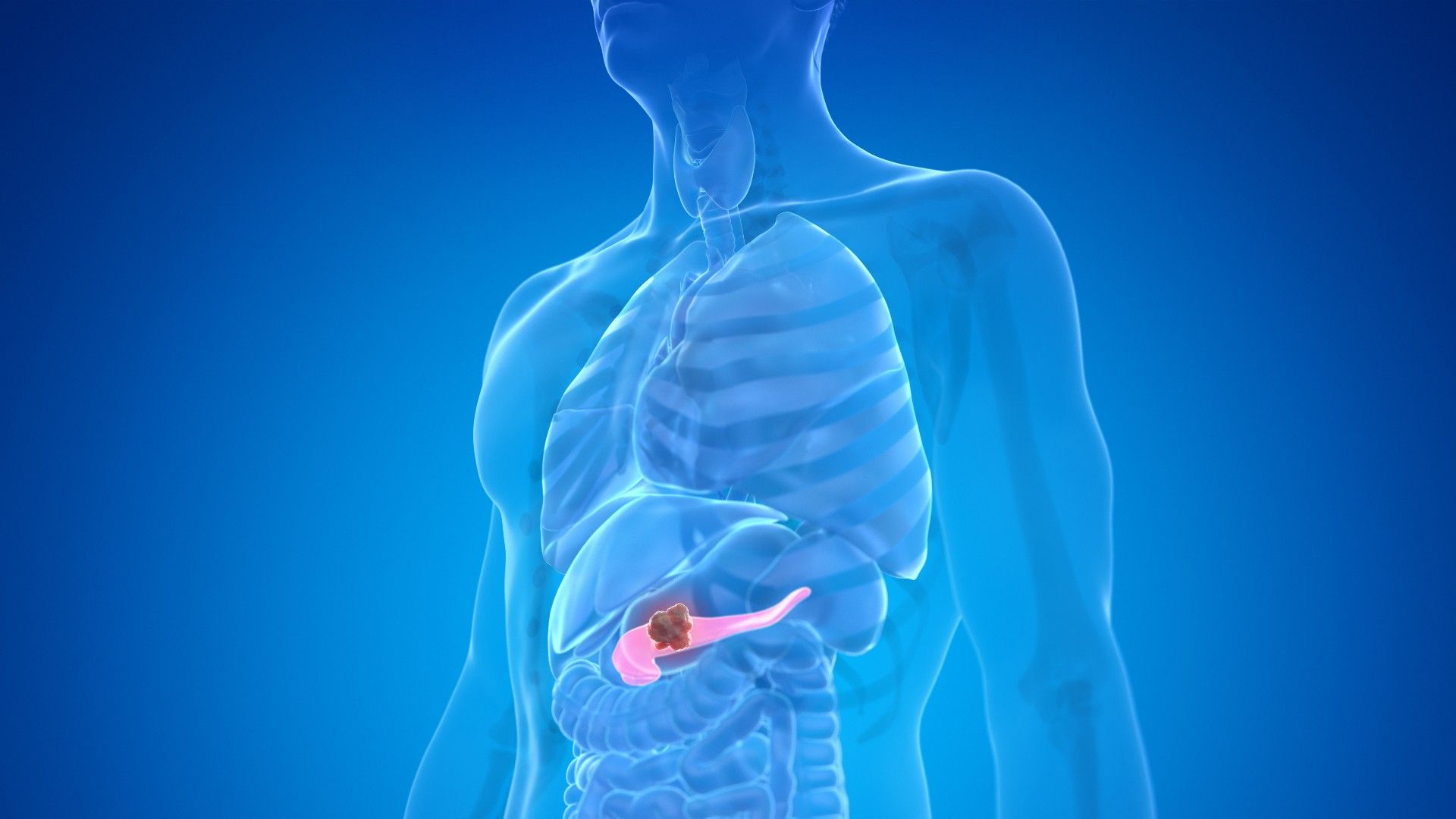
Pancreatic cancer is one of the deadliest malignancies, often diagnosed too late for effective treatment. Recent breakthroughs in artificial intelligence (AI) have demonstrated that deep learning models can identify subtle patterns in routine CT scans—patterns invisible to the human eye—that signal the presence of pancreatic cancer up to three years before conventional diagnosis. This guide walks you through the process of how such an AI model operates, from data collection to clinical interpretation, offering a clear, actionable overview for healthcare professionals, researchers, and tech enthusiasts. By understanding each step, you can appreciate the potential and limitations of this cutting‑edge tool in transforming early cancer detection.

What You Need
- High‑quality CT scan dataset – A large, annotated collection of abdominal CT scans, including both healthy patients and those later diagnosed with pancreatic cancer. The dataset must cover a timeline of at least three years before diagnosis to train the AI to spot early indicators.
- Pre‑trained deep learning model – A convolutional neural network (CNN) architecture, such as a modified U‑Net or ResNet, specifically designed for medical image analysis. The model should have been previously trained on a diverse set of CT scans to recognize pancreatic tissue and subtle anomalies.
- Computing resources – A workstation or cloud environment with a powerful GPU (e.g., NVIDIA A100 or V100) and sufficient RAM (at least 32 GB) to process volumetric CT data. Software frameworks like TensorFlow or PyTorch are required.
- Data preprocessing tools – Image normalization scripts, segmentation algorithms to isolate the pancreas region, and augmentation libraries to increase dataset diversity.
- Clinical validation team – Radiologists and oncologists to interpret AI outputs and correlate them with patient outcomes, ensuring the model’s predictions are clinically meaningful.
Step‑by‑Step Process
Step 1: Collect and Annotate CT Scans
Begin by assembling a comprehensive retrospective dataset of abdominal CT scans from a known cohort. For each scan, record the patient’s eventual diagnosis and the exact date of clinical confirmation. The key is to include scans taken three, two, and one year before that diagnosis, as well as control scans from patients who remained cancer‑free. Radiologists must annotate the scans with bounding boxes around the pancreas and any suspicious lesions, even if they were originally dismissed as benign. This ground truth is essential for supervised learning.
Step 2: Preprocess and Normalize Images
Raw CT scans vary in resolution, contrast, and orientation. Standardize all images to a uniform voxel size (e.g., 1.0 mm isotropic) using linear interpolation. Apply windowing to highlight soft tissues (window width ~400 HU, window level ~40 HU). Normalize pixel intensities to [0,1] or Z‑score. Segment the pancreas region automatically with a pre‑trained organ segmentation model to reduce noise from surrounding organs. Finally, augment the dataset with random rotations, flips, and elastic deformations to improve model robustness.
Step 3: Train the AI Model
Split the preprocessed dataset into training (70%), validation (15%), and test (15%) sets. Load the deep learning model with randomly initialized or pre‑trained weights. Optimize using a loss function such as binary cross‑entropy for cancer/non‑cancer classification, or a combination of focal loss and Dice loss for segmentation. Use a batch size that fits your GPU memory (e.g., 4–8 volumes). Train for 100–200 epochs with early stopping based on validation AUC. Monitor training curves to avoid overfitting; if necessary, increase dropout or L2 regularization.
Step 4: Validate the Model’s Performance
After training, evaluate the model on the held‑out test set. Calculate sensitivity, specificity, positive predictive value, and area under the receiver operating characteristic curve (AUC). Most importantly, measure how early the model flags cancer compared to the true diagnosis date. In the original study, the AI detected signs up to three years prior. Subset analysis by stage and by patient demographics ensures the model works across diverse populations. If performance is insufficient, revisit data preprocessing, model architecture, or training hyperparameters.
Step 5: Integrate into Clinical Workflow
Once validated, deploy the AI model as a decision support tool. For new incoming CT scans, the model runs automatically and outputs a risk score. If the score exceeds a threshold (e.g., ≥0.7), it flags the case for radiologist review. The radiologist examines the original scan with the AI’s attention heatmap overlays, which highlight the most suspicious regions. A positive finding triggers additional imaging (e.g., MRI or endoscopic ultrasound) or biopsy, while a negative result maintains standard follow‑up. Establish a feedback loop: every confirmed cancer or false positive should be added to the training dataset for continuous improvement.
Tips for Success
- Prioritize data quality. The AI’s accuracy hinges on the richness of the training data. Include scans from multiple institutions, scanners, and protocols to prevent overfitting to a single machine.
- Collaborate with domain experts. Radiologists and pathologists must validate the model’s predictions—AI flags subtle texture changes that may be invisible even to experienced clinicians.
- Plan for regulatory approval. If you intend to use this model in a clinical setting, prepare for FDA or CE marking. Document every step: data sources, model architecture, training parameters, and validation results.
- Manage false positives. Early detection may increase unnecessary biopsies. Set specificity high (e.g., 95%) to maintain clinical trust, even at the cost of some sensitivity.
- Integrate with electronic health records. Automate the flow of CT scans to the AI and the AI’s output back into the patient record to minimize radiologist workload.
- Communicate uncertainty. Always present AI predictions as probabilities, not certainties. Educate clinicians on interpreting the model’s confidence scores.
- Prepare for evolution. AI models can drift as imaging technology changes. Establish a retraining schedule—annually or whenever new CT protocols are introduced.
By following these steps, you can replicate the breakthrough that promises to transform pancreatic cancer from a near‑certain death sentence into a treatable condition. The journey from research to routine clinical use requires rigorous validation, but the potential reward—saving lives through early intervention—is immense.
Related Articles
- From Constipation Relief to Kidney Protection: A Surprising Medical Breakthrough
- Breakthrough: Common Constipation Drug Shows Promise in Slowing Kidney Disease
- Artificial Eggshell Breakthrough: A Key Step Toward Reviving Extinct Birds?
- 10 Ways Medicare's New ACCESS Payment Model Is Revolutionizing AI in Healthcare
- Revolutionary Injectable Material Repairs Damaged Organs from Within the Bloodstream
- AstraZeneca's Hypertension Drug Gets FDA Green Light: Key Questions Answered
- Workplace Mental Health Crisis: Over Half of U.S. Employees Report Crying on the Job in Past Month
- Cannabis After 65: A Step-by-Step Safety Guide Based on Stanford Expert Warnings